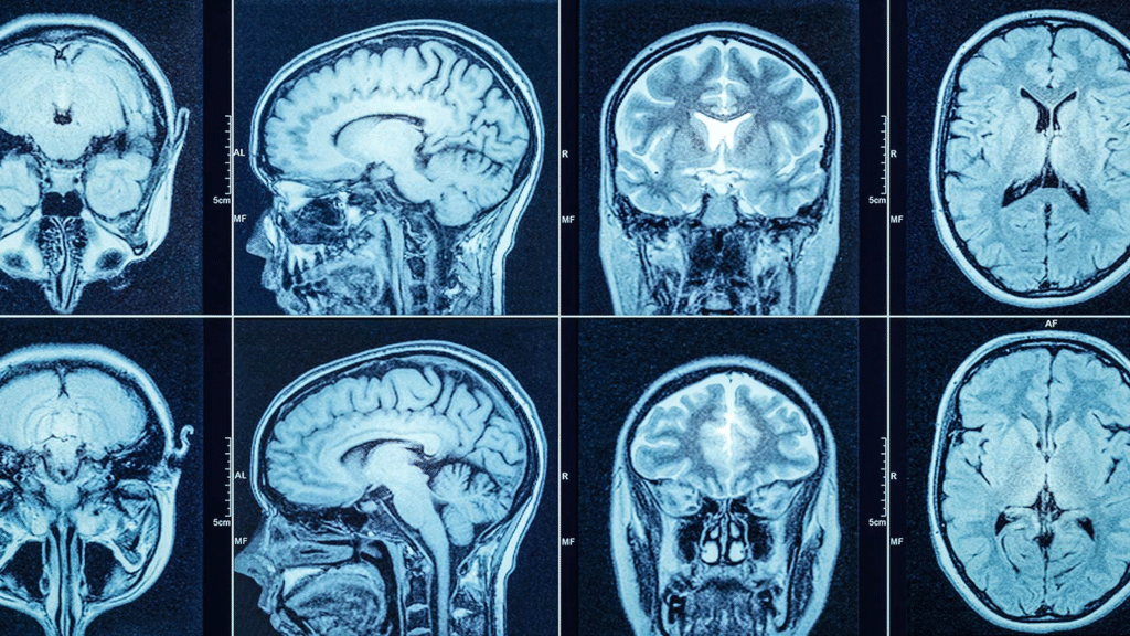
A Computed Tomography (CT) scan, also known as a CAT scan, is a powerful diagnostic tool that uses X-ray beams and computer processing to generate detailed cross-sectional and 3D images of the body. Developed in the late 1960s and refined through the 1970s, CT earned a Nobel Prize in 1979 for its revolutionary impact on medical imaging. Since the development, CT scans have become indispensable in modern medicine and beyond.
What Is a Computed Tomography (CT) X-ray?
A CT scan is a medical imaging technique that uses motorized X-ray sources rotating around a patient to collect data from multiple angles. The CT scan system processes the collected data to produce ‘slices’ of the body. It provides far more detail than conventional X-rays and can reveal information about bones, organs, blood vessels, and soft tissues.
Also read: People Screening Equipment: All You Need to Know
How Does a CT Scan Work?
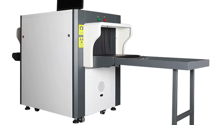
A CT scanner consists of:
- Gantry: Circular frame housing the X-ray tube and detectors that spin around the patient.
- X-ray Tube & Detectors: Emit and detect X-rays, converting them into electrical signals.
- Patient Table & Slip Rings: Moves through gantry continuously (in helical CT); slip rings prevent cable entanglement.
- Computer & Console: Process data, reconstruct images, and display results.

Narrow X-ray beams rotate around the body while the table advances, capturing multiple slices that are stacked or displayed in 3D for comprehensive analysis.
Types of Computed Tomography Scans Explained
- Sequential CT (Step‑and‑Shoot): Uses step-wise table movement; slower scanning.
- Spiral or Helical CT: Continuous table motion with rotating X-ray tube; faster and more efficient.
- Electron Beam Tomography (EBT/Ultrafast CT): Utilizes electronically swept electron beam for rapid heart imaging.
- Dual‑Energy CT: Employs two X-ray energy levels via dual or switched sources for enhanced tissue differentiation.
- CT Perfusion Imaging: Measures blood flow, volume, and transit time using contrast—especially useful in stroke and heart studies.
- PET‑CT (Positron Emission Tomography + CT): Combines PET functional imaging with CT anatomical detail—crucial in cancer detection and staging.
Also read: Trace Detection Equipment: Where are they Used?
Why Are CT Scans Used?

CT scans are key tools for diagnosing and managing numerous conditions:
- Detecting tumors, internal bleeding, trauma, and bone injuries.
- Planning biopsies and surgical interventions.
- Evaluating organs like the brain, lungs, heart, abdomen, and blood vessels.
CT Scan With or Without Contrast: What’s the Difference?
Using contrast media enhances the visibility of blood vessels and soft-tissue structures:
- IV Contrast (Iodine-based): Highlights vascular structures like coronary arteries.
- Oral or Rectal Contrast (Barium-based): Used for gastrointestinal tract imaging.
Contrast helps improve diagnostic clarity, though it may cause temporary effects such as flushing, metallic taste, headache, or nausea.
Also read: How Does Baggage and Parcel Inspection Equipment Work | Full Guide
Who Performs a Computed Tomography Scan?
Radiographers or radiology technologists operate CT scans, positioning patients, managing scanner settings, and ensuring image quality. Radiologists—medical doctors specialized in imaging—interpret the scans and issue diagnostic reports.
CT Scan Procedure: What to Expect
Preparation
- Fasting required for IV contrast (3 or 4 hours).
- Remove metal objects, piercings, and wear a gown.
- Disclose allergies and kidney conditions.
During the Scan
- Lie still on a motorized table that slides into the gantry.
- Technologists control the scan from a separate room but maintain communication.
- Each rotation lasts a few seconds, with the overall duration varying based on the type of examination and contrast.
After the Scan
- Monitor for contrast reactions if used (e.g. rash, difficulty breathing).
- There’s no need for a specific recuperation period; proceed with your regular activities unless instructed otherwise.
Are Computed Tomography Scans Safe?
- CTs use ionizing radiation, posing a small increase in lifetime cancer risk.
- CT scan professionals take safety precautions for pregnant patients and children, opting for alternative imaging, such as magnetic resonance imaging (MRI) or ultrasound when possible .
- Contrast agents carry allergy and kidney injury risks but are generally well tolerated.
Also read: Where to Buy the Best Explosives and Narcotics Detection Systems in Nigeria
Computed Tomography Scans Beyond Medicine: Other Applications
Industrial and scientific applications of CT scanners include:
- Industrial inspection: flaw detection, metrology, reverse engineering.
- Airport security: 3D baggage scanning for explosives and weapons.
- Geological & Paleontological research: internal imaging of rocks and fossils.
- Cultural heritage: analyzing artifacts and scrolls non-invasively.
- Microbiology: visualizing sub-micron structures in fungi.
- Timber industry: detecting defects like knots in sawmill operations.
Also read: What are Advanced Security Screening Products & Solutions?
Limitations and Challenges of CT Scans
- Image artifacts: Things like metal implants or prostheses (such as hip replacements) can cause bright streaks or dark shadows on CT images. These are called artifacts and they can make it hard to see the body clearly.
For example, metal in the body can confuse the CT scanner and create streaks that block important details. Even though there are methods to reduce these effects, they don’t always work perfectly and can sometimes cause new problems. Scientists are working on better ways to fix this—like a new algorithm that reduces metal artifacts without needing to know what kind of metal is in the implant. Such an approach could help doctors plan radiation treatment more accurately for people with metal implants. - Radiation exposure and repeated scans increase risk.
- Cost and availability: Equipment can be expensive and not always accessible.
Advancements in Computed Tomography Imaging
Recent developments in computed tomography (CT) imaging are transforming its capabilities. These emerging advancements include:
- AI-enhanced reconstruction to boost image quality and reduce artifacts.
- Faster and higher-resolution scanners improving temporal and spatial clarity.
- Expansion of industrial and non-medical CT applications, powered by innovations in hardware and software.
FAQs About Computed Tomography X‑rays
- What does a CT scan detect that other tests might miss?
Beyond standard X-rays, a CT scan can reveal fine details in soft tissue, complex fractures, and vascular issues. - Is a CT scan painful?
No. It’s non-invasive and painless. However, lying still may cause discomfort. - How long does a CT scan take?
A CT scan can take anywhere from a few minutes to an hour. It is quicker without contrast and takes longer with it. - Can I eat before a CT scan?
Yes—unless contrast is used. Then fasting for ~3 hours is required. - Are CT scans covered by insurance?
Coverage varies by region and provider; generally covered when deemed medically necessary. - Is there an age limit or risk group for CT scans?
Special care is taken with children and pregnant women to minimize radiation impact
Conclusion
A CT scan provides unmatched internal imaging through detailed slices and 3D reconstructions. It’s a cornerstone in medical diagnostics. Its applications range from treating trauma and stroke to detecting cancer and planning surgical procedures, and it also plays crucial roles in industries, security, conservation, and research.
Follow us on X (formerly Twitter), @Logic_sss, for more security guides and updates.
References
- www.hopkinsmedicine.org – Computed Tomography (CT) Scan
- www.nibib.nih.gov – Computed Tomography (CT)
- en.wikipedia.org – CT scan


